Why does my dentist want to take X-rays?
Radiographs/x-rays are a valuable diagnostic tool when seeing a dentist to check your current oral condition. There are several types of x-rays used in a dental setting the most common of which include:
Bitewings
Taking routine x-rays (every two years) allows the practitioner to check areas that are not otherwise visually accessible. This x-ray shows the crowns of both the upper and lower teeth but does not extend down to the root tips of either.
This may benefit the patient in several ways:
- They highlight areas that need attention or more focused cleaning such as demineralisation of the enamel between the teeth. By pointing out areas such as this a practitioner aims to prevent any progression of disease in these areas. This allows practitioners to watch suspicious areas rather than immediately filling small areas of demineralisation on teeth that may otherwise be controlled with good oral hygiene.
- Series of bitewings provide a timeline of disease, changes are watched and tracked over time if there were a sudden change in an otherwise stable mouth a time frame of the disease progression can aid in narrowing down the cause or change in lifestyle that has affected the patient. This allows the practitioner to ask more targeted questions and give more practical and personalised advice relating to a patient’s oral health.
- Prevent larger fillings or more major works from being necessary; in some areas of the mouth or due to the structure of some people’s enamel, decay will not be apparent until it has become quite severe. Having x-rays allow the dentist to advise on decay when it is in it’s early stages hopefully before more major work is necessary.
These radiographs usually include a device or a plate with a wing/tab on which to bite onto once placed on the inside of the teeth. These single image dental x-rays are very low exposure, 2x images are approximately less than 0.0025mSv of radiation which is equivalent to less than half a day of natural background radiation.
In a stable mouth, bitewings can be taken every two years but for patients with high decay or rapid changes dentists may advise that yearly bitewings would be beneficial.
Periapicals
This is another in chair dental x-ray however it will target a specific tooth showing clinicians the tooth from the tip of the crown to the very apex of the root of the tooth.
The benefit of having a periapical before treatment is that:
- The practitioner is able to support a diagnosis with radiographic evidence. Some conditions such as a root fracture, a dental abscess or internal resorption are not necessarily able to be seen by visually inspecting the mouth and do require a periapical x-ray to properly diagnose these conditions.
- Shows root formation and attachment to bone. Everyone is individual and there are some people that have roots that are shaped differently to others or some teeth that may fuse to the bone. Before treatment it is important for practitioners to know these details before commencing with some procedures that may be affected by this.
- To check the quality of treatment. Dentists may take a periapical during or after invasive treatment such as a filling or root treatment to be sure that the quality of the work is high and provides the best treatment possible for the patient.
These radiographs are not taken routinely and are generally requested when a patient or practitioner has a concern regarding a particular tooth. They are the same low dose radiation exposure as a single bitewing x-ray.
OPG or Panoramic
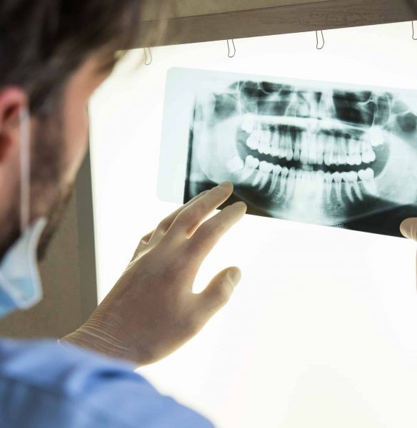
This 2-dimensional dental x-ray shows the full mouth including all the teeth the jaw and the palate giving a good overview of the general health of the mouth. An approximate
These x-rays can track many things and can benefit a patient by:
- Showing eruption patterns in children/adolescents with mixed dentition or for checking the eruption of the third molar (wisdom teeth).
- Give an overview of the periodontium (bone that surrounds the teeth). Using these x-rays clinicians can check for any periodontal disease that affects the bone before damage has become too severe.
- Check for fractures in the underlying structures of the jaw or palate.
- An OPG will quite often be used to assess for the adequacy of the bone before referring for a more detailed x-ray when looking at implants.
Clinicians are more discerning when referring for OPG/panoramic x-rays as their radiation exposure is higher than a single x-ray at approximately 0.014mSv which is equivalent to less than 3 days’ worth of natural background radiation exposure.
Other x-rays may include:
- A Lat Ceph – to check the profile view of the teeth and bone structure, usually used when referring for orthodontic treatment.
- A CBCT (Cone beam CT) – to check for the viability of implants or for information that cannot be obtained from single two-dimensional images such as root resorption or precise positioning of teeth.
- TMJ x-rays – check the jaw joint for any abnormalities in the case of pain, severe clicking or crunching of the joint.

